
Primary CNS Lymphoma in AIDS


Comments:
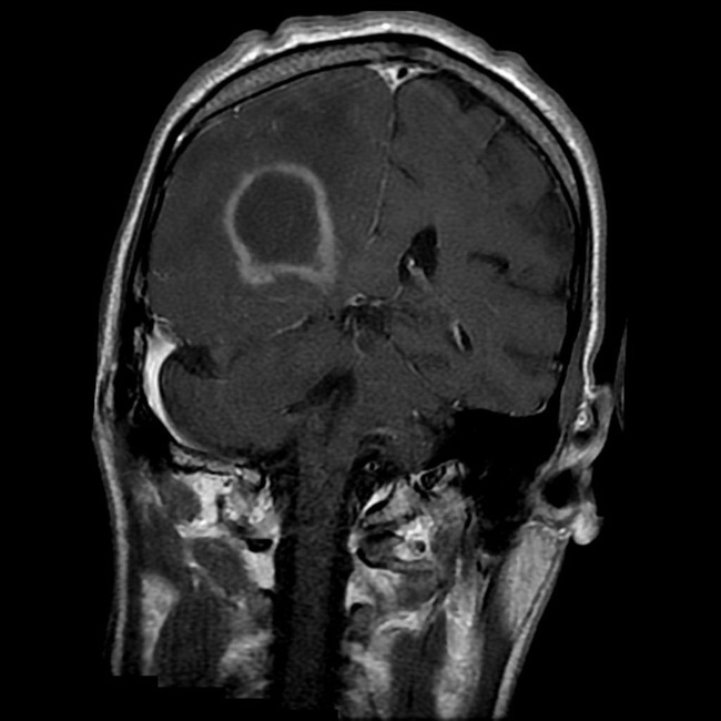
This HIV+ adult male with low CD4 count presented with headaches. An MRI with contrast showed a mass in the parietal lobe that is peripherally enhancing with central hypodense areas (consistent with necrosis). There was considerable surrounding vasogenic edema. The mass was biopsied. Sections showed an angiocentric infiltrate of large atypical lymphoid cells with the following immunohistochemical profile: POSITIVE for CD20, Ki67, P53, and EBV. They were negative for CD10 and BCL6. A few CD3+ reactive T-cell were present in the background. Case courtesy of A.Prof Frank Gaillard, Radiopaedia.org. From the case rID: 5374 The incidence of primary CNS lymphoma has increased in recent years due to the increased number of immunocompromised patients (organ transplant recipients, HIV infection, etc.). PCNSL develops in 2% to 12% of AIDS patients. The patients are younger with a striking male predominance in contrast to PCNSL in immunocompetent patients.