
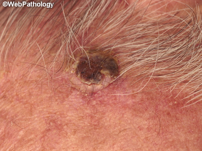
Keratoacanthoma : Clinical Presentation


Comments:
Keratoacanthoma : Clinical Variants - continued from previous slide. Keratoacanthoma centrifugum marginatum: Rare keratoacanthomas can become quite large and multinodular. They show peripheral expansion and central healing. The lesions heal slowly over 6-12 months or persist for years, expanding gradually and causing considerable tissue destruction. Keratoacanthoma in Muir-Torre Syndrome: Keratoacanthoma may develop within a pre-existing nevus sebaceus or be a manifestation of Muir-Torre syndrome. Fergusson-Smith Syndrome: An autosomal dominant condition seen largely in Scottish population that consists of multiple self-healing keratoacanthomas. The lesions may number into hundreds and present in childhood or early adulthood. They occur on sun-exposed skin, are slow to heal, and cause considerable scarring. Grzybowski Syndrome: The patients develop large numbers of eruptive papular lesions that can be pruritic or painful on sun-exposed skin. Subungual Keratoacanthoma: A rare variant that arises from the nail matrix and is more destructive than the usual keratoacanthomas. It usually involves thumb or forefinger and presents with pain, swelling, and erythema. There may be destruction of the underlying bone. Treatment consists of curettage.