
Melanotic Neuroectodermal Tumor of Infancy


Comments:
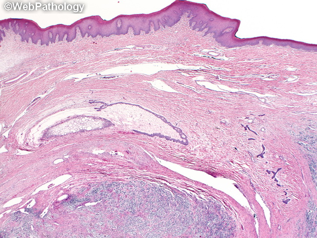
Melanotic Neuroectodermal Tumor of Infancy (MNTI) is tumor of neural crest origin with dual neuroblastic and epithelioid differentiation. It has been known by a variety of names, including melanotic progonoma, melanotic adamantinoma and retinal anlage tumor. It usually presents within the 1st yr. of life as a cystic, radiolucent, rapidly growing, locally destructive tumor involving the maxilla. Rare cases have involved other cranial bones, brain, epididymis, uterus and mediastinum. The tumor produces disfiguring facial deformity and blue-gray to black mucosal pigmentation depending upon the amount of melanin produced. Rare cases are associated with elevated levels of urinary vanillylmandelic acid. This specimen is from a 9 month old boy with a 4-month history of a rapidly enlarging, protruding mass in his oral cavity causing dysphagia. The mass involved his left maxilla along the alveolar ridge and bulged into his maxillary sinus causing displacement of teeth. The resected mass was firm, gray-white with patchy brown-black pigmentation. Intact oral mucosa overlying the tumor is seen at the top. The tumor is at the bottom of the image. The diagnosis was MNTI. Histologic features are shown in slides 42-46.