
Medulloblastoma : MRI
slide 1 of 23


Comments:
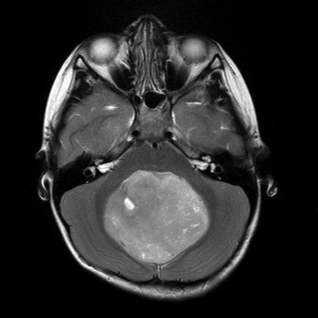
MRI of the brain of a 6 year old child showing a large mass located centrally within the posterior fossa. It has high T2 signal (as seen in this image) and showed only patchy contrast enhancement. No normal vermis can be identified and the mass is likely arising from it, completely effacing the fourth ventricle. The radiographic findings are consistent with a medulloblastoma. The diagnosis was confirmed on biopsy. Case produced with permission, courtesy of Dr. Frank Gaillard. Radiopaedia. Complete case is here.
slide 1 of 23