
Inverted Papillary Uroth. CA, High-Grade : Differential


Comments:
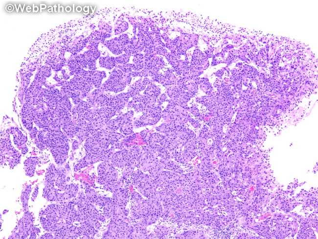
Differential Diagnosis: Urothelial carcinomas with predominantly inverted growth pattern vs inverted urothelial papilloma. Features favoring Inverted Urothelial Papilloma: smooth overlying surface with no or minimal exophytic component, circumscribed lesion with a smooth base, thin uniform anastomosing cords and trabeculae within lamina propria, microcysts or pseudoglandular spaces, no cytologic atypia, peripherally palisading basal cells, centrally streaming spindle or ovoid cells, minimal amount of simple fibrovascular stroma, and no obvious stromal infiltration or stromal response (desmoplasia). Low Ki-67 labeling index. Negative for UroVysion FISH test. Low incidence of LOH. Features favoring Urothelial Carcinomas with Inverted Growth: prominent exophytic component, thickened irregular cords and large nests with solid foci within lamina propria (seen here), cytologic atypia with increased mitotic activity, and destructive stromal invasion (for invasive tumors). High Ki-67 labeling index. Positive for UroVysion FISH test. Features not present include: peripheral palisading and central streaming of urothelial cells, superficial umbrella cells, and microcystic or pseudoglandular spaces.