
Inverted Papillary Uroth. CA, High-Grade : Microscopic
Home
 Genitourinary
Urinary Bladder
Inverted Urothelial Neoplasms
Inverted Papillary Uroth. CA, High-Grade : Microscopic
Genitourinary
Urinary Bladder
Inverted Urothelial Neoplasms
Inverted Papillary Uroth. CA, High-Grade : Microscopic

slide 73 of 80


Comments:
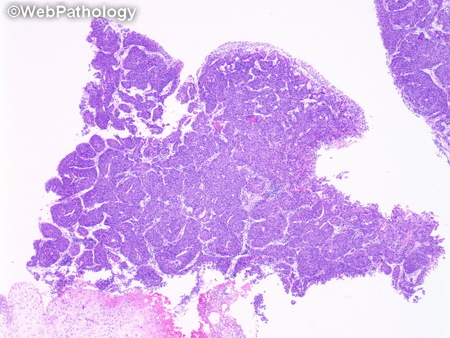
Microscopic Features: This image from a transurethral resection specimen shows inverted papillary urothelial carcinoma, high-grade, invasive. There are thick, irregular, anastomosing trabeculae invading the lamina propria. At high magnification, these tumors show overtly malignant cells with enlarged or irregular, hyperchromatic nuclei and prominent nucleoli. Additional features of malignancy include apoptosis, necrosis, neovascularization, discohesiveness, and increased mitotic activity with atypical mitoses. Features usually absent include peripheral palisading and central streaming of urothelial cells, superficial umbrella cells, and microcystic or pseudoglandular spaces.
slide 73 of 80