
Inverted Papillary Urothelial Neoplasm of LMP : Microscopic
Home
 Genitourinary
Urinary Bladder
Inverted Urothelial Neoplasms
Inverted Papillary Urothelial Neoplasm of LMP : Microscopic
Genitourinary
Urinary Bladder
Inverted Urothelial Neoplasms
Inverted Papillary Urothelial Neoplasm of LMP : Microscopic

slide 48 of 80


Comments:
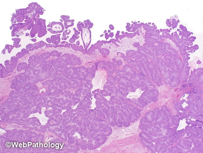
Microscopic Features of Inverted Papillary Urothelial Neoplasm of LMP:
Unlike the thin, uniform cords and trabeculae of inverted urothelial papilloma, the neoplastic urothelial cells are organized in large, markedly thickened and irregular cords and trabeculae as well as solid nodules within the lamina propria. The peripheral palisading and central streaming of urothelium, seen in IUP, may or may not be present. A surface papillary component is commonly seen.
slide 48 of 80