
Inverted Urothelial Papilloma

slide 36 of 80


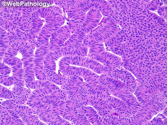
Comments:
The urothelial cells are arranged in orderly cords and trabeculae and show no cytologic atypia. Focal degenerative type atypia is seen in some cases. Mitotic activity is not increased.
slide 36 of 80