
Mature Cystic Teratoma : Microscopic

slide 53 of 183


Comments:
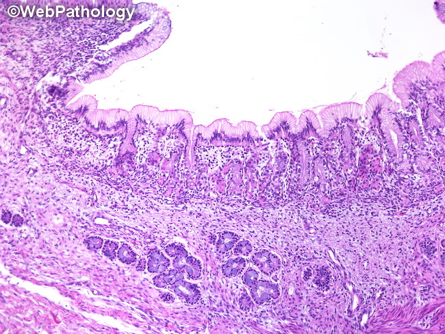
MICROSCOPIC FEATURES (continued from the previous image): The cyst wall in mature cystic teratomas is lined by various types of epithelia, including skin, respiratory mucosa, intestinal epithelium, and even follicular thyroid cells. The image shows gastric mucosa lining the cyst. Certain tissues such as keratin and fat may cause secondary changes such as foreign body reaction and lipogranulomatous response. In patients with anti-NMDA receptor encephalitis-associated ovarian teratomas, there is prominent lymphoid infiltration of neuroglial tissues, with formation of germinal centers. (continued in the next image)
slide 53 of 183