
Mature Cystic Teratoma : Microscopic


Comments:
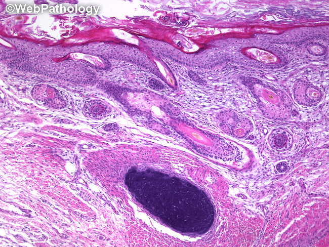
MICROSCOPIC FEATURES (discussed in the next several images): By definition, mature teratomas contain mature somatic tissues that are histologically similar to their eutopic counterparts but are haphazardly laid out within the tumor. The tissues are derived from two or all three germ cell layers as follows:ECTODERMAL STRUCTURES (present in 100% of cases) - mature epidermis with adnexal structures (seen here), neuroectodermal elements (glia, ependyma, cerebellar tissue, whorls of meningothelial cells, Wagner-Meissner corpuscles); MESODERMAL ELEMENTS (93% of cases) - bone, cartilage (seen here), adipose tissue, smooth muscle, and glomeruloid vascular proliferation; ENDODERMAL ELEMENTS (71% of cases) - respiratory, gastrointestinal, salivary glands, thyroid tissue, and rarely, prostate, pituitary, parathyroid, adrenal tissue, and various types of neuroendocrine cells. (continued in the next image)