
Dysgerminoma : Microscopic


Comments:
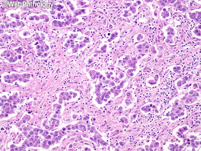
Dysgerminomas resemble seminomas microscopically. They are composed of monomorphic tumor cells arranged in diffuse sheets, trabeculae, cords, or nests separated by delicate fibrous septae. There may be pseudoglandular spaces or poorly-formed tubules due to discohesion among tumor cells. The fibrous septae contain lymphocytes (T cells), plasma cells, eosinophils, and sometimes histiocytes. In some cases, there may be well-formed lymphoid follicles with germinal centers. About 20% of cases show granulomatous response consisting of epithelioid granulomas. The inflammatory component may be prominent enough to obscure the neoplastic cells. The polygonal tumor cells have well-defined cell membranes, clear or lightly eosinophilic cytoplasm, large vesicular nuclei, one or two prominent nucleoli, and brisk mitotic activity. The nuclei often have an angulated or squared-off appearance. The cytoplasmic clarity is due to glycogen content. About 5% of dysgerminomas have syncytiotrophoblastic cells, usually in a perivascular location. Such cases show modest elevations of serum hCG levels. This feature does not change the prognosis. Choriocarcinoma must be excluded by thorough sampling. Rare cases show luteinized stromal cells. Microcalcifications may be seen in cases arising in gonadoblastomas.