
Mucinous Borderline Tumor : Microscopic


Comments:
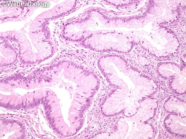
The image shows mucinous borderline tumor (MBT) - intestinal type. The glands are lined by a single layer of mucinous columnar epithelium with basally-located hyperchromatic nuclei. Besides glandular structures, the epithelial lining can show multilayering, tufting as well as branching papillary structures. The papillae are either stroma-free or have thin fibrous cores. The lining epithelium is usually admixed with goblet cells (as seen in this image), neuroendocrine cells as well as rare Paneth cells. There is mild to moderate cytologic atypia in the form of nuclear enlargement and hyperchromasia with occasional prominent nucleoli (best seen in the large gland in the lower left quadrant in this image). The appearance resembles a hyperplastic or adenomatous colonic polyp. High-grade nuclear features are not seen. Mitotic activity is variably increased. There is no destructive stromal invasion. The cytologic atypia should be seen in at least 10% or more of the tumor for it to be designated MBT.