
Synovial Sarcoma : Case History

slide 7 of 78


Comments:
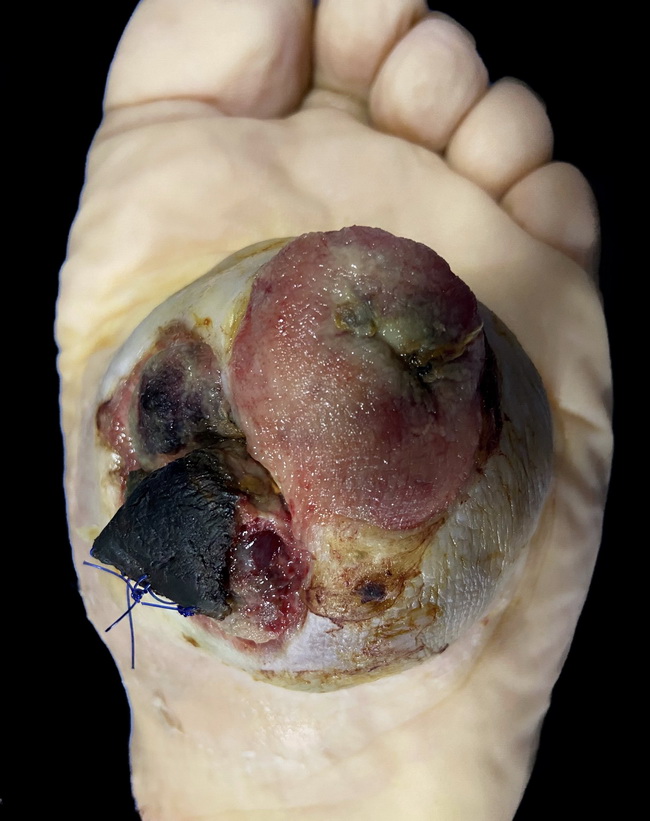
This foot amputation specimen is from a 70 y/o female with a 3-month history of a foot nodule. Microscopic features were classic for a monophasic synovial sarcoma. Immunohistochemical markers showed the following: Positive for TLE1, EMA, and BCL2; Negative for actin, desmin, and S-100 protein. As discussed in slide 1, almost 85-95% of synovial sarcomas arise in the extremities, especially around knee or ankle joints. Image courtesy of: Luis Soler, MD, Mexico; used with permission.
slide 7 of 78