
Synovial Sarcoma : Monophasic


Comments:
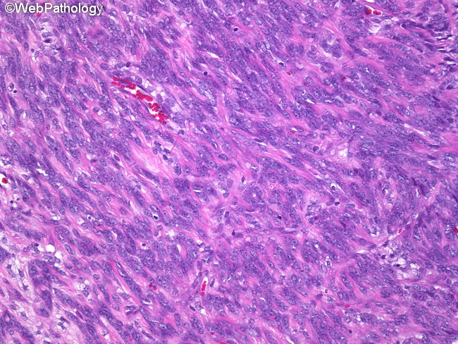
Monophasic synovial sarcoma, as the name implies, consists of only one component. It is usually a monophasic fibrous type (the most common subtype of synovial sarcoma) composed purely of spindle cells which can mimic fibrosarcoma, malignant peripheral nerve sheath tumor, or other sarcomas. The sarcomatous areas are hypercellular and consist of monotonous fibroblast-like spindle cells with plump nuclei that may be arranged in whorls or intersecting fascicles. Hemangiopericytomatous vascular pattern (stag-horn vessels), focal nuclear palisading, and abundant mast cells are commonly seen. The intervening stroma can show myxomatous change, hyalinization, calcification (20% of cases), or osseous metaplasia. Some cases show prominent cystic change. Some monophasic fibrous synovial sarcomas lack obvious epithelial component but may show clusters of cells with pale cytoplasm and an epithelioid morphology. A monophasic epithelial synovial sarcoma (or more appropriately called epithelial-predominant synovial sarcoma) does occur; however, this diagnosis is rarely made and can be reliably established only with the help of molecular genetic or cytogenetic studies. Several entities that enter into the differential diagnosis must be ruled out, including metastatic carcinoma, melanoma, adnexal tumors, epithelioid sarcoma, and epithelioid malignant peripheral nerve sheath tumor.