
Synovial Sarcoma : Biphasic


Comments:
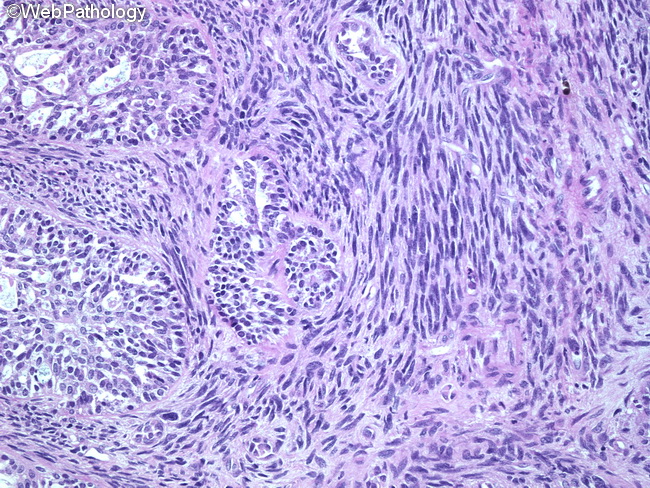
Biphasic synovial sarcoma consists of epithelial and spindle cell components in varying proportions. Epithelial areas: They resemble adenocarcinoma and are composed of columnar or cuboidal cells arranged in solid cords, nests, or gland-like formations with granular or eosinophilic secretions. Some areas show papillary or villous structures lined by cuboidal or flattened epithelium. The epithelial cells have round to oval vesicular nuclei, abundant pale cytoplasm, and distinct cellular borders. Focal squamous metaplasia may be seen. Spindle component: The sarcomatous areas are hypercellular and consist of monotonous fibroblast-like spindle cells with plump nuclei. The spindle areas resemble fibrosarcomas and may show whorls, intersecting fascicles, hemangiopericytomatous vascular pattern (stag-horn vessels), focal nuclear palisading, and abundant mast cells. The intervening stroma can show myxomatous change, hyalinization, calcification (20% of cases), and osseous metaplasia. Some cases show prominent cystic change. Hemorrhage and necrosis are more common in poorly-differentiated tumors.