
Endometriosis of Ureter

slide 28 of 39


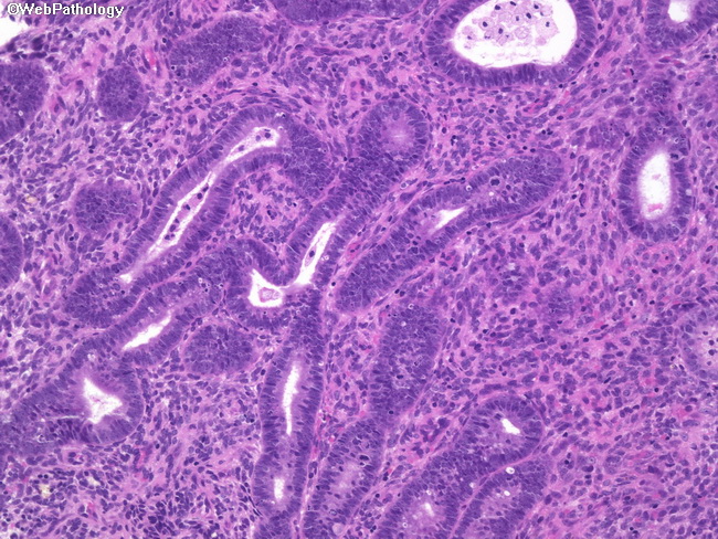
Comments:
This ureteral biopsy is from a 52 y/o female who presented with intermittent gross hematuria. Work-up revealed right-sided hydronephrosis and a 4 cm pelvic mass. Ureteroscopy showed a polypoid, inflammatory-appearing lesion in the distal ureter which turned out to be endometriosis (shown here). Benign endometriotic gland are surrounded by typical stroma. The pelvic mass underwent CT-guided needle biopsies which showed endometrioma.
slide 28 of 39