
Renal Angiosarcoma : Immunostains


Comments:
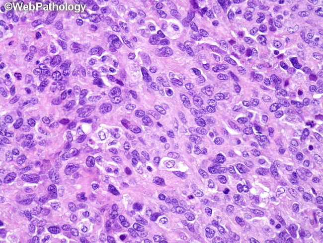
Immunohistochemistry: Immunohistochemical stains are essential in distinguishing renal angiosarcomas from other renal neoplasms. Angiosarcomas are negative for epithelial markers like AE1/AE3, EMA, and CAM5.2, although focal positivity may be seen in a small percentage of cases with epithelioid morphology. They are also negative for RCC, CK8/18, CD10, S-100 protein, Melan-A, and HMB-45. On the other hand, they are positive for endothelial markers like CD31, CD34, FLI-1, Factor VIII-related antigen, and ERG. This image shows a high-grade angiosarcoma that was primary in the kidney. The tumor cells have epithelioid morphology with abundant eosinophilic cytoplasm and large vesicular nuclei with punctate nucleoli. The presence of better differentiated areas with anastomosing vascular channels lined by atypical endothelial cells (noted elsewhere in the specimen) as well as strong immunoreactivity for CD31 and CD34 confirmed the diagnosis.