
Ischemic Fasciitis : Microscopic

slide 5 of 12


Comments:
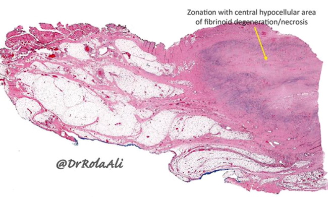
Microscopic Features of Ischemic fasciitis: Ischemic fasciitis shows a zonal pattern which is obvious at low magnification. There is a central area of fibrinoid necrosis surrounded by a zone of vascular proliferation resembling granulation tissue (darker blue/purple area at the periphery of the lesion in this image) and ganglion-like myofibroblasts. Image courtesy of: Rola Ali, MD, FRCPC, Associate Professor of Pathology, Kuwait University, Kuwait; used with permission.
slide 5 of 12