Ischemic Fasciitis : Differential Diagnosis


Comments:
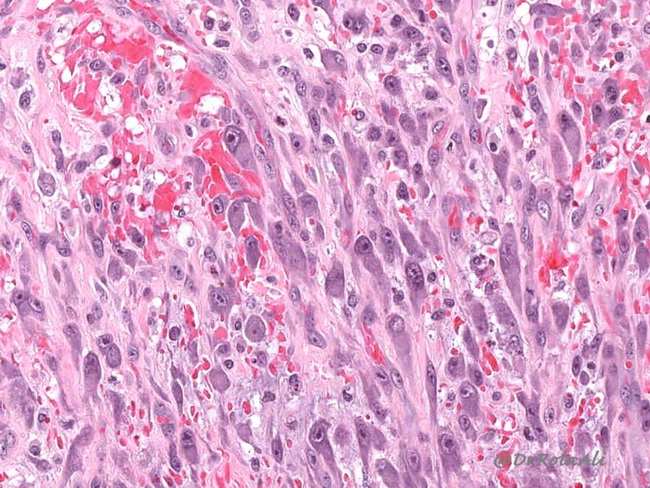
Ischemic fasciitis can resemble the following entities: Epithelioid Sarcoma: The presence of central necrosis in ischemic fasciitis with proliferation of atypical cells can mimic epithelioid sarcoma. However, epithelioid sarcoma occurs in the distal extremities of young patients, and exhibits prominent cytoplasmic eosinophilia, cytokeratin immunoreactivity, and loss of INI1 expression. Myxoid Liposarcoma: In myxoid areas of ischemic fasciitis, one may see multivacuolated muciphages which can mimic atypical lipoblasts. However, true lipoblasts are not seen. Moreover, ischemic fasciitis also lacks the plexiform vasculature of myxoid liposarcomas. Myxofibrosarcoma: It lacks the zonation of ischemic fasciitis. Other reactive features of ischemic fasciitis, including smudgy hyperchromatic nuclei, fat necrosis, hemosiderin deposition, and fibrin thrombi are also absent in myxofibrosarcoma. The image shows large ganglion-like myofibroblasts in a case of ischemic fasciitis. Similar cells are also seen in nodular fasciitis, proliferative fasciitis, and proliferative myositis. Image courtesy of: Rola Ali, MD, FRCPC, Associate Professor of Pathology, Kuwait University, Kuwait; used with permission.


