
Lipoblastoma : Clinical Features


Comments:
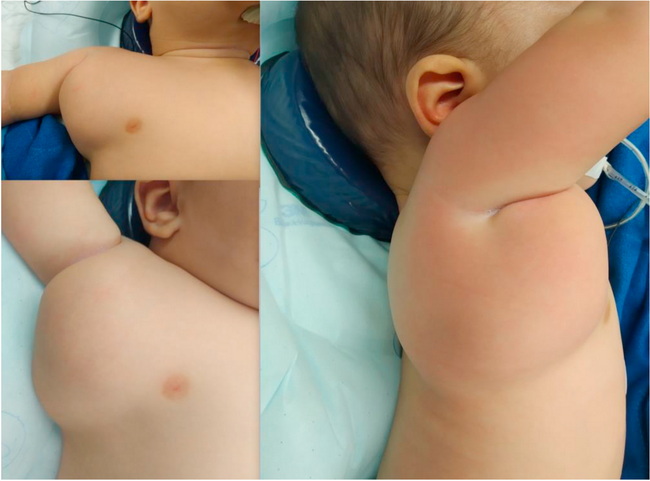
Clinical Features of Lipoblastoma (continued from the previous image): The superficial lesions (lipoblastomas) can clinically resemble an ordinary lipoma or a hemangioma. They present as a painless, circumscribed, subcutaneous mass ranging in size from 1 to 15 cm. The deep-seated tumors (lipoblastomatosis) are ill-defined and often infiltrate skeletal muscle. They may become quite large and compress vital structures. The tumors in the head and neck region may compress airways producing stridor. Thoracic tumors may spread to spinal canal producing hemiparesis or quadriparesis. Colonic and mesenteric tumors can result in intussusception and volvulus. A small group of lipoblastoma patients have disorders of the central nervous system, including seizures, autism, developmental delay, congenital abnormalities, and Sturge-Weber syndrome.About this image: Lipoblastoma presenting as a rapidly growing, 8 x 6 cm, soft, subcutaneous mass in the axilla in a 6 month old boy. The diagnosis was confirmed by histological examination of the resected specimen. Image source: Shrestha A.L., Kabs C., Wenzel R., Hosie S. Childhood Lipoblastoma. Journal of Pediatric Surgery Case Reports. Vol. 63, Dec. 2020, 101677; Used under Creative Commons Attribution Non-commercial NoDerivatives 4.0 International License.