
Chondroid Lipoma : Immunohistochemistry


Comments:
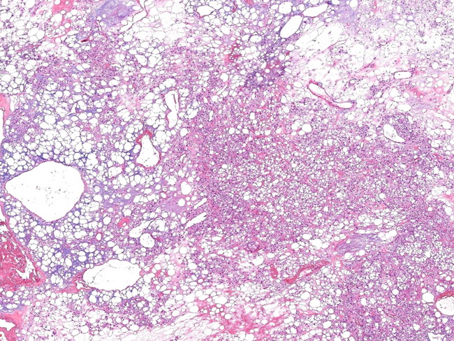
Immunohistochemistry: Chondroid lipomas show positivity for S-100 protein (in lipoblasts) and may show focal immunoreactivity for CD68 (vacuolated cells) and cytokeratin. EMA is negative. Cyclin D1 expression has been found in one study. The tumor is negative for HMB-45, SMA, MSA, and GFAP. Ki-67 proliferation index is <1%. Ultrastructurally, the tumor cells show a range of differentiation from prelipoblasts and chondroblasts to lipoblasts, preadipocytes and mature adipocytes.This low magnification view shows an admixture of mature adipocytes, lipoblasts, and hibernoma-like cells with eosinophilic granular cytoplasm. Image courtesy of: Andy Zhang, MD, Dept. of Pathology, Rushan People's Hospital, Rushan City, Shandong Province, China.