
Chondroid Lipoma : Microscopic Features

slide 7 of 14


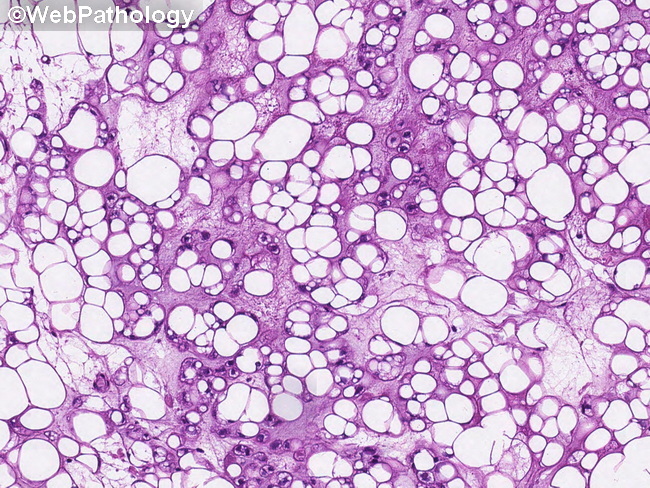
Comments:
Microscopic Features (continued from the previous image): The amount of mature adipose tissue in chondroid lipoma is variable and can be focal or extensive. The background matrix is myxoid and may show zones of hyalinization. There may be areas of inflammation, hemorrhage, hemosiderin deposition, dystrophic calcification, or even metaplastic bone formation. The vascularity is prominent and consists of thick-walled vessels as well as cavernous spaces. Some cases have osteoclast-like multinucleated cells.This image shows an admixture of mature adipocytes, uni- or multivacuolated lipoblasts and small round tumor cells with chondroid features.
slide 7 of 14