
Chondroid Lipoma : Microscopic Features

slide 6 of 14


Comments:
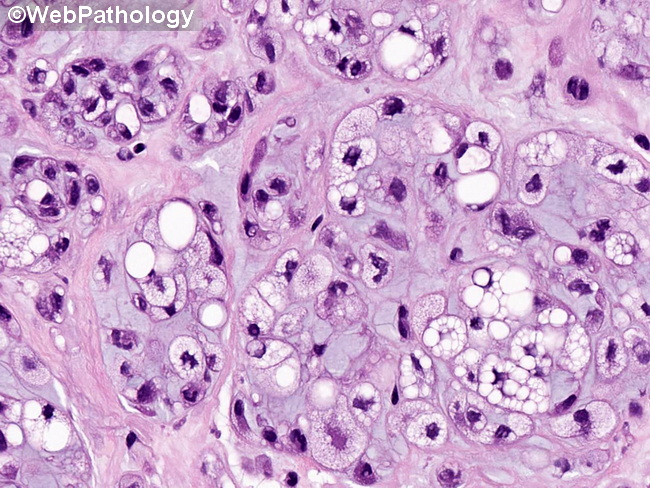
Microscopic Features (continued from the previous image): The rounded tumor cells are arranged in sheets, nests and cords in a background of myxoid or hyalinized chondroid stroma. The presence of lacuna-like spaces around small tumor cells in a myxohyaline background creates resemblance to cartilage. There is variable amounts of mature fat admixed with uni- or multivacuolated lipoblasts and hibernoma-like cells with eosinophilic granular cytoplasm. The cells have uniform oval or bean-shaped nuclei with even chromatin and inconspicuous nucleoli. There is no pleomorphism and mitotic activity is low.
slide 6 of 14