
Chondroid Lipoma : Differential Diagnosis


Comments:
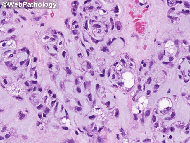
Differential Diagnosis of Chondroid Lipoma (continued): Spindle cell lipomas - arise superficially in the dermis or subcutis; location on the back or posterior neck; lack chondroid features; contain ropey collagen fibers; carry deletion of 13q or 16q.Soft tissue chondroma - occurs in hands and feet; contains true hyaline cartilage and multinucleated giant cells; lacks fat vacuoles.Myoepithelial tumors - are more superficial, usually have ductal or epithelial foci and lack lipoblasts. The immunostains for keratins, S-100 protein, smooth muscle actin and GFAP are positive. This image of chondroid lipoma shows mostly small round cells with eosinophilic cytoplasm in a myxoid matrix. The cells have uniform hyperchromatic nuclei. A few multivacuolated lipoblasts are also present. There is close resemblance to extraskeletal myxoid chondrosarcoma.