
Intramuscular Lipoma

slide 31 of 40


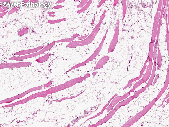
Comments:
Grossly, intramuscular lipomas appear as areas of yellow fat partially replacing the muscle tissue. Microscopically, there are mature adipocytes diffusely infiltrating the skeletal muscle. The involved muscle fibers may show some atrophy; however, there are no lipoblasts or cells with atypical nuclei. Atypical lipomatous tumor/well-differentiated liposarcoma (ALT/WDL) must be excluded by thorough sampling. If there is any doubt, the use of FISH to demonstrate the absence of MDM2 amplification is helpful in excluding the diagnosis of ALT/WDL. The prognosis is excellent if the tumor is completely resected.
slide 31 of 40