
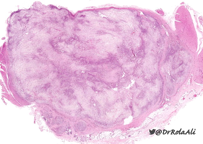
Nodular Fasciitis : Microscopic

slide 7 of 36


Comments:
The appearance of nodular fasciitis at low magnification depends upon the age of the lesion. In the earlier phases, it appears bluish with a loosely textured, feathery pattern due to abundant myxoid matrix. Lesions of longer duration show dense hyaline fibrosis and formation of microcysts. In cystic nodular fasciitis, smaller cysts coalesce to form a large central cyst. Image courtesy of: Rola Ali, MD, FRCPC, Associate Professor of Pathology, Kuwait University, Kuwait; used with permission.
slide 7 of 36