
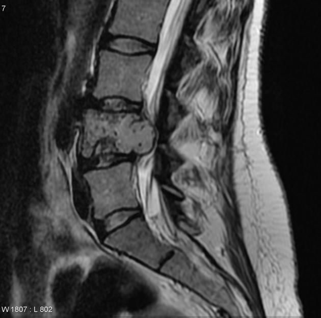
Chondrosarcoma : Spine

slide 8 of 60


Comments:
This MRI scan (T2) from a 34 y/o woman shows an expansile, lytic lesion involving L4 vertebra. The tumoor has eroded posterior cortex but is confined by the posterior longitudinal ligament with no extension into the soft tissues. The patient underwent complex resection of L4 vertebral body including left side posterior elements and associated soft tissues, adjacent intervertebral discs, inferior body of L3 and superior body of L5. Microscopic examination showed a clear cell chondrosarcoma. Case produced with permission, courtesy of Dr. Frank Gaillard. Radiopaedia. Complete case is here.
slide 8 of 60