
Metastatic GIST


Comments:
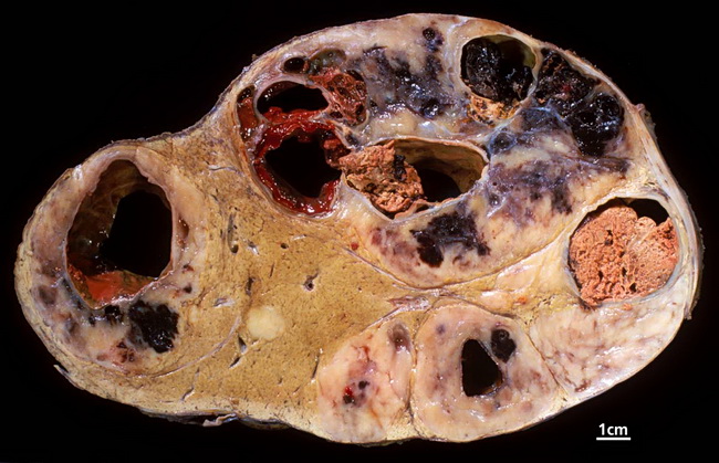
Gastrointestinal stromal tumors and leiomyosarcomas are the most frequent sarcomas to metastasize to the liver. With GIST, FNA smears show relatively monomorphic spindle cells arranged singly or in small aggregates. The background may show myxoid stroma. With epithelioid GIST, the differential diagnosis includes melanoma, carcinoma, and neuroendocrine tumors. Needle core biopsies will show sheets of plump epithelioid cells with eosinophilic cytoplasm. Classic GISTs show high vascularity, paranuclear vacuoles, variably myxoid or hyalinized stroma, and sometimes skenoid fibers (extracellular collagen globules). Immunohistochemical analysis shows positivity for CD117, DOG1, CD34, and vimentin. SMA, cytokeratins, and S-100 are variably positive. Desmin is negative. This gross specimen photograph of a liver slice is from a young female patient with history of GIST of stomach. Two years after gastrectomy she presented with liver metastases. There are several tumor nodules with hemorrhage and necrosis. Image copyright: pathorama.ch.