
Liver Hemangioma : Additional Images
Home
 Gastrointestinal
Liver
Liver Tumors & Tumor-like Lesions - I
Liver Hemangioma : Additional Images
Gastrointestinal
Liver
Liver Tumors & Tumor-like Lesions - I
Liver Hemangioma : Additional Images

slide 25 of 63


Comments:
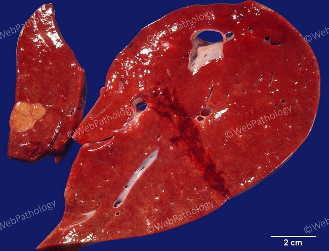
Incidental liver findings at autopsy. The large slice shows a hemangioma. The smaller slice shows a peribiliary gland hamartoma (bile duct adenoma). Peribiliary gland hamartomas are small (usually < 1 cm), solitary, subcapsular yellow-white lesions that may be mistaken for metastases. Microscopically, they consist of small tubular structures with abortive lumens, embedded in a fibrous stroma.
slide 25 of 63