
Unicystic Ameloblastoma : Intro


Comments:
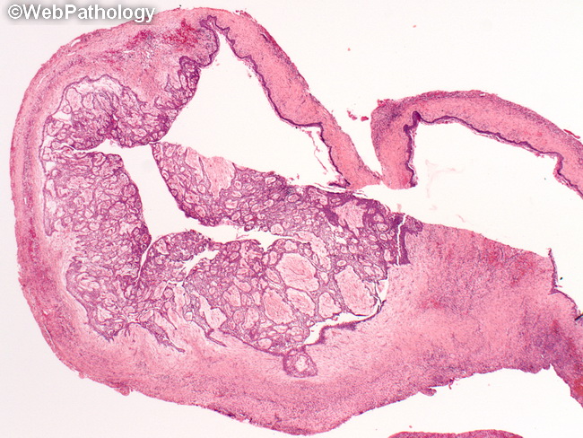
Unicystic ameloblastoma has been considered as a separate entity given its distinct clinical, radiologic, and pathologic features. It comprises 15% to 20% of all ameloblastomas. It may originate de novo as a neoplasm as well as by neoplastic transformation of epithelial lining of a benign odontogenic cyst. It occurs in a younger age group than conventional solid/cystic ameloblastoma. The median age is 2nd decade of life (vs around 35 years for conventional ameloblastoma). The vast majority of cases involve posterior mandible and present as painless expansile lesion. On plain radiographs, it appears as a circumscribed radiolucent lesion associated with the crown of an unerupted mandibular 3rd molar. Clinically and radiologically, unicystic ameloblastoma is difficult to distinguish from dentigerous, primordial, radicular, or residual cyst. The diagnosis becomes apparent only after histopathologic examination of the cyst wall.