
Hyaline-Vascular Castleman Disease
Home
 Hematopathology
Lymph Node (Non-Hematopoietic)
Lymphadenopathies - I
Hyaline-Vascular Castleman Disease
Hematopathology
Lymph Node (Non-Hematopoietic)
Lymphadenopathies - I
Hyaline-Vascular Castleman Disease

slide 56 of 69


Comments:
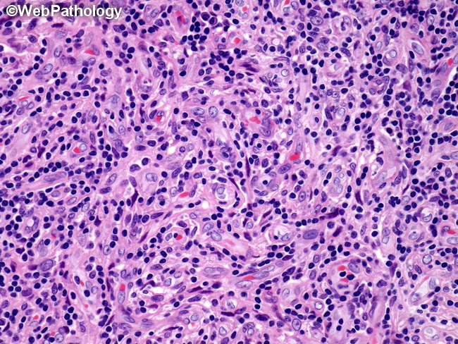
Microscopic Features of Hyaline-Vascular Castleman Disease (HVCD) (continued): The interfollicular region is greatly expanded by proliferation of high endothelial venules (as shown here) and a mixed cell population of plasma cells, eosinophils, plasmacytoid dendritic cells (CD123+), and TdT+ T-cells. A stroma-rich variant of HVCD has been described in which there are prominent vascular or actin-positive myoid elements in the interfollicular region and may even form so-called angiomyoid proliferative lesions.
slide 56 of 69