
Kimura Disease : Microscopic Features


Comments:
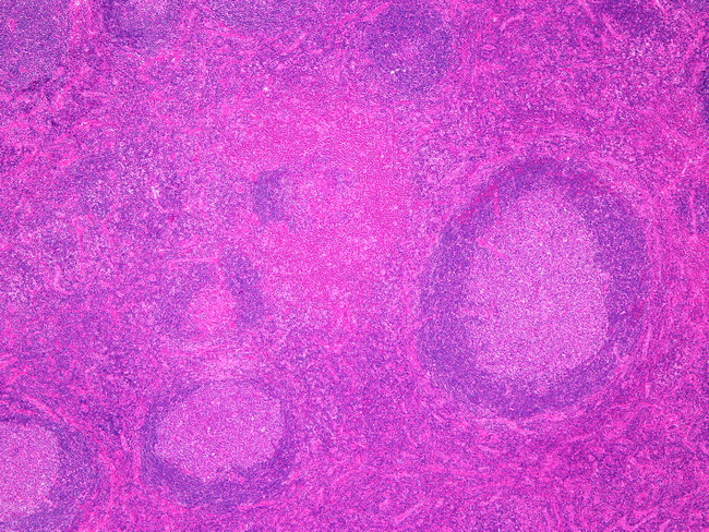
Microscopic Features of Kimura Disease: The affected lymph nodes are enlarged but the nodal architecture is largely preserved. There are hyperplastic follicles with reactive germinal centers and a well-defined mantle zone. The germinal centers are hypervascular and show polykaryocytes (Warthin-Finkeldey cells) and deposits of proteinaceous material. A prominent feature is extensive eosinophilic infiltrate, often with formation of eosinophilic microabscesses. Additional features include proliferation of postcapillary venules, perivenular and stromal sclerosis, and increased number of mast cells and IgG4+ plasma cells. This image shows several hyperplastic follicles. The interfollicular zone is expanded by eosinophilic infiltrate and post-capillary venules. An eosinophilic microabscess in seen just above the center of the image. Image courtesy of: @Patholwalker