
Sclerosing Pneumocytoma (Hemangioma)

slide 5 of 17


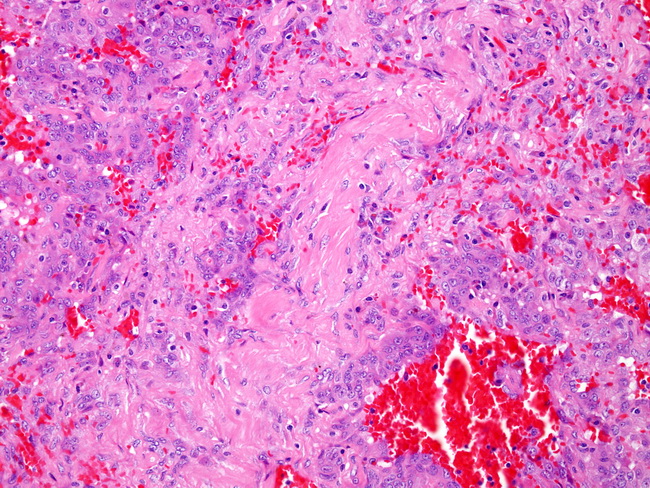
Comments:
The epithelioid cells are present, forming variably sized ectatic spaces filled with red blood cells typical of the hemorrhagic pattern. The epithelioid cell is one of two cell types that may be seen in sclerosing hemangioma (the other cell type is discussed later). The sclerotic foci punctuate the surrounding hemorrhagic pattern.
slide 5 of 17