
Inverted Urothelial Papilloma : Differential Diagnosis


Comments:
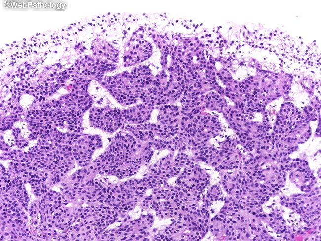
Differential Diagnosis of Inverted Urothelial Papilloma (IUP) (continued from the previous image): Urothelial carcinoma with inverted pattern (shown here) has thick, irregular columns of malignant-appearing cells. The trabeculae often transition to solid areas. The presence of exophytic papillary areas, invasion of muscularis propria, necrosis, marked cytologic atypia, or increased mitotic activity support the diagnosis of inverted urothelial carcinoma. In contrast, the cords and trabeculae in IUP have uniform and orderly arrangement. The trabeculae show centrally streaming spindle cells and peripherally palisading darker basal cells. There is no cytologic atypia or increased mitoses. Muscularis propria is not involved.