
Peripheral Ossifying Fibroma : Differential Diagnosis
Home
 Head & Neck
Oral Cavity, Oropharynx & Neck
Soft Tissue Tumors of Oral Cavity
Peripheral Ossifying Fibroma : Differential Diagnosis
Head & Neck
Oral Cavity, Oropharynx & Neck
Soft Tissue Tumors of Oral Cavity
Peripheral Ossifying Fibroma : Differential Diagnosis

slide 15 of 29


Comments:
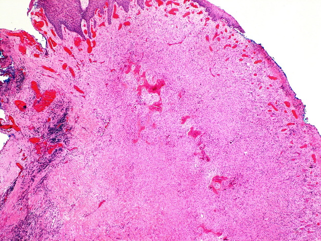
Differential Diagnosis of peripheral ossifying fibroma (POF) includes irritation fibroma, pyogenic granuloma, and peripheral giant cell granuloma. This image of a POF shows a bland spindle cell proliferation under squamous mucosa that is intact on the left and ulcerated on the right. The deeper aspects of the lesion show osteoid matrix. Image courtesy of: Dr. Ibrahim Zardawi; used with permission.
slide 15 of 29