May 2017
Neurofibroma
Reviewer(s): Dharam M. Ramnani, MD
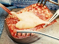
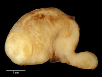
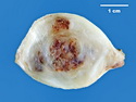
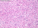
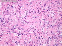
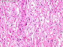
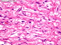
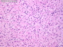
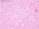
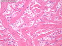
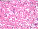
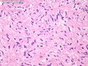
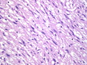
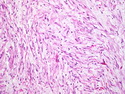
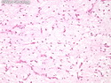
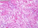
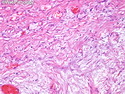
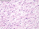
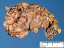
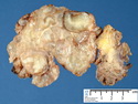
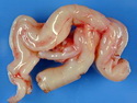
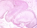
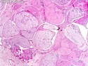
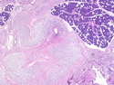
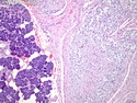
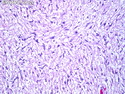
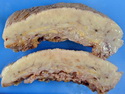
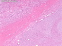
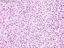
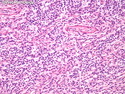
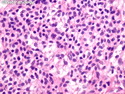
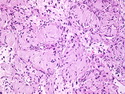
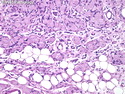
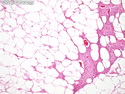
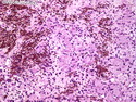
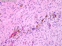
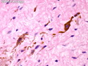
Neurofibroma is a benign nerve sheath tumor arising in the peripheral nervous system. It may be sporadic or associated with Neurofibromatosis 1. Based on the growth pattern, it is subdivided into localized, plexiform, and diffuse types. Localized Neurofibroma: It usually occurs as solitary, superficial cutaneous tumor in individuals who do not have Neurofibromatosis 1 (NF1). They are much more common than those associated with NF1. They present as slow-growing painless lesions in the dermis or subcutis in individuals 20-30 years of age. Plexiform Neurofibroma (PF): They develop in early childhood and are virtually diagnostic of NF1. They can be found in superficial as well deep locations converting the involved large nerve trunks and nerve roots into a thickened tortuous mass resembling 'a bag of worms.' They can cause massive enlargement of the entire extremity, hypertrophy of the long bones, and redundancy and hyperpigmentation of skin. PF may display increased cellularity as well as nuclear atypia. Such lesions are at a greater risk of malignant transformation. Diffuse Neurofibroma (DF): DF involves the head and neck region in children and young adults and is seen sporadically as well as in NF1. It is poorly-circumscribed and causes plaque-like thickening of the skin and subcutaneous tissues. References: 1) Enzinger & Weiss's Soft Tissue Tumors, Sixth Edition, 2014; p. 793-813. 2) Rosai & Ackerman's Surgical Pathology, Tenth Edition, 2011; p. 2132-35.