
Solid Pseudopapillary Tumor : Microscopic


Comments:
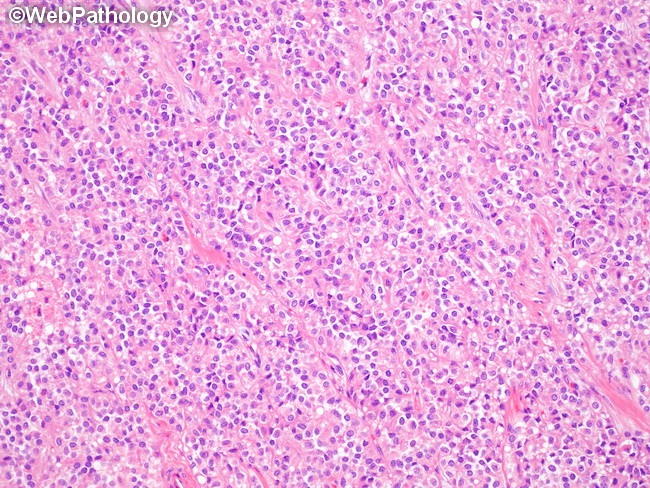
Microscopic Features of Solid Pseudopapillary Tumor (SPT) of Pancreas: SPT shows varied histologic patterns, including solid, pseudopapillary and hemorrhagic-cystic areas. The solid areas at the periphery of the tumor consist of poorly-cohesive sheets of small, uniform polygonal cells admixed with delicate vasculature. The cells have light pink to clear cytoplasm, indistinct nucleoli, and frequent longitudinal nuclear grooves. True gland formation is not seen. Mitotic activity is not increased. A tumor with a predominant solid pattern may resemble Acinar Cell Carcinoma (which is seen mostly in males and is trypsin and chymotrypsin positive) and Well Differentiated Pancreatic Neuroendocrine Tumor (keratin and chromogranin A positive). SPT is immunoreactive for vimentin, CD10, CD56, synaptophysin, α1 anti-trypsin, progesterone receptors, and shows nuclear accumulation of β-catenin.