
Trichoepithelioma


Comments:
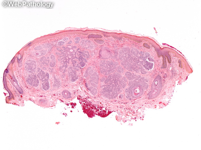
Trichoepithelioma is composed of nests of basaloid cells with horn cysts lying in the dermis. The tumor cells have minimal cytoplasm, large hyperchromatic nuclei and show peripheral palisading. The appearance is reminiscent of basal cell carcinoma. The overlying epidermis may be normal, hyperkeratotic or thinned with loss of rete ridges. The tumor lobules are invested by a sheath of dense fibrous connective tissue. Another frequently seen feature is the formation of dense round or oval aggregates of fibroblastic cells referred to as papillary mesenchymal body. The papillary mesenchymal body may indent into the lobules of basaloid cells. The absence of this feature in basal cell carcinoma is a helpful distinguishing feature. Uncommonly seen features include ulceration of overlying epidermis, foreign body giant cell reaction to keratin, calcification, and amyloid deposits. Trichoepitheliomas arising in extra-facial sites are likely to be well-circumscribed, show little or no keratin cyst formation and composed of irregularly shaped lobules of basaloid cells in a background of dense connective tissue.