
Hidradenoma Papilliferum

slide 69 of 126


Comments:
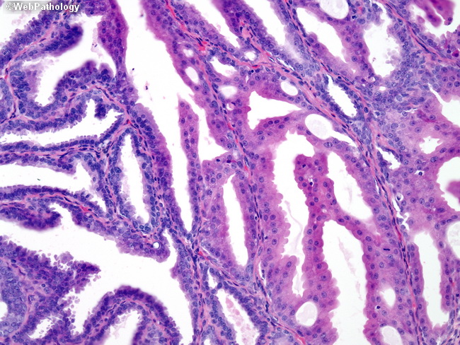
Some cases may show a varied appearance throughout the lesion. In the photomicrograph above, some of the cells (right) show marked apocrine changes. These cells contain abundant, pink cytoplasm and prominent apical snouts.
slide 69 of 126