
Trichobezoar : Diagnosis & Treatment


Comments:
Discussion continued from the previous slide: Diagnosis of Bezoars: Clinical history is extremely valuable in arriving at the diagnosis. The history of the type and the amount of foods and medications ingested should be obtained. A history of previous bezoar or gastric surgery, if available, is helpful. On physical examination, a palpable abdominal mass may sometimes be present. The patient may have halitosis due to putrefaction of the stomach contents. Patients with trichobezoar may show evidence of trichotillomania in the form of baldness and patchy hair loss pattern. Imaging studies & Endoscopy: A plain abdominal film may outline the bezoar. In contrast studies, the bezoar is seen as a large filling defect in the stomach. Upper endoscopy is most helpful in making the diagnosis. Phytobezoars appear as a dark green, brown, or black mass of amorphous material. Trichobezoars are firm to hard and black in color. Pharmacobezoars show pills or pill fragments embedded in amorphous material. Treatment: Small bezoars can be treated conservatively with a liquid diet, a prokinetic agent to improve gastric motility and speed up gastric emptying, and chemical dissolution with cellulase (given as tablets). Small phytobezoars can be dissolved with nasogastric lavage and even with carbonated sodas (like Coca Cola). Larger bezoars can be fragmented during endoscopy and the fragments removed via mouth or pushed into small intestine. If endoscopic therapy is unsuccessful or if there is a complication such as bleeding or perforation caused by bezoar, operative intervention is done. Gastric bezoars are removed via gastrostomy and small intestinal bezoars are removed by enterotomy. Preventing recurrence of bezoar is equally important. The underlying causes should be addressed. This may include avoidance of high-fiber and non-digestible foods, using prophylactic cellulase capsules, prokinetic drugs for patients with impaired gastric motility, and psychiatric treatment for patients with trichobezoar. Reference:
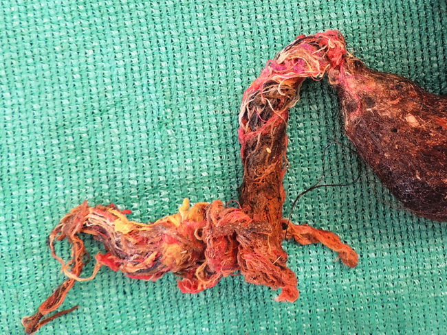
Feldman, M., Friedman, L. S., & Brandt, L. J. (2016). Sleisenger & Fordtran's Gastrointestinal & Liver Disease - 10th Edition. Philadelphia, PA. Elsevier Saunders. Vol 1; Chapter 27; p. 434-435. This image shows extension of trichobezoar (same case as the previous image) into the duodenum (Rapunzel syndrome). Case courtesy of Dr. Sanjay D. Deshmukh (Professor of Pathology) and Dr. Jayant M. Gadekar (Professor & Head of Surgery), Dr. Vithalrao Vikhe Patil Foundation's Medical College and Hospitals, Ahmednagar, India.