
Fibrolamellar Carcinoma of Liver

slide 55 of 65


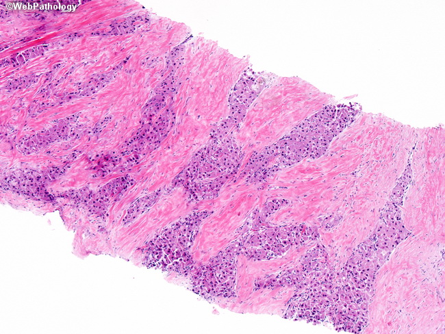
Comments:
FLC is distinguished from conventional HCC by unique histologic features, namely: parallel fibrous lamellae separate groups of large, polygonal, densely eosinophilic tumor cells, which contain abundant mitochondria. The fibrotic stroma resembles that of the scirrhous pattern of conventional HCC but, in FLC, is present throughout the tumor in dense, parallel lamellae. The image shows FLC in a needle biopsy of liver.
slide 55 of 65