
Lipoma : Imaging

slide 5 of 40


Comments:
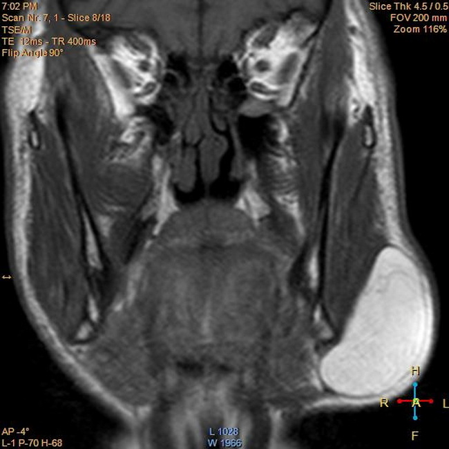
Imaging Studies: Both CT and MRI are excellent imaging modalities for the diagnosis of lipomas. This MRI (coronal T1-weighted image) is from a 60 y/o male who presented with a painless swelling on the left side of neck. The left submandibular region shows an oval shaped well-defined mass. It exhibits high signal in T1, T2-weighted images. There is no bony or soft tissue infiltration. It was confirmed to be a lipoma. Case courtesy of Ahmed Abdrabou, Radiopaedia.org. From the case rID: 22468
slide 5 of 40