
Primary Breast Angiosarcoma : Microscopic


Comments:
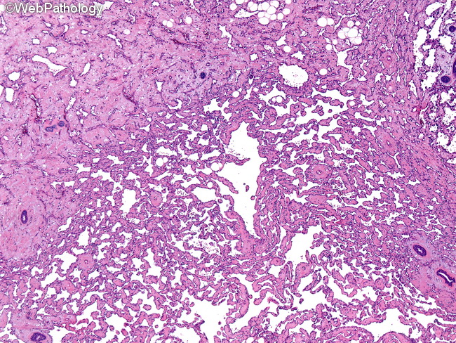
The microscopic features of breast angiosarcomas are similar to those seen elsewhere and include a broad spectrum. There is vasoformative growth, papillary endothelial growth, solid growth, and capillary hemangioma-type growth. The tumor cells may be typical flattened endothelial type or have plump, spindled, or epithelioid phenotype. The nuclear atypia ranges from minimal to severe. Areas of necrosis and hemorrhage resulting in blood lakes are seen in high-grade tumors. Grading of Breast Angiosarcomas: Breast angiosarcomas are classified into three grades based on growth patterns, degree of nuclear atypia, and mitotic activity; however, the prognostic value of tumor grade is not certain. Multiple histologic grades often coexist within a single tumor. The overall histologic grade is determined on the resected specimen and not in biopsies to prevent undergrading. Grade I (low) angiosarcomas of the breast have well-defined, irregularly-shaped, anastomosing vascular channels (as seen here) lined by minimally atypical flattened endothelium. The vascular channels are variably dilated or have compressed slit-like lumina. They diffusely infiltrate within the breast parenchyma and fat and surround the terminal duct lobular units. The diagnosis of malignancy is rendered on the basis of infiltrative growth pattern. Nuclear atypia is minimal, mitotic activity is not significantly increased, and papillary endothelial growth is not a prominent feature.