
Metaplastic Meningioma

slide 42 of 60


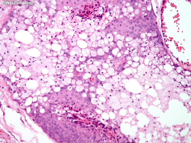
Comments:
This is a higher magnification of the previous picture illustrating areas of xanthomatous metaplasia. Xanthomatous appearance is due to the accumulation of lysosomes or small lipid vacuoles within meningothelial cells. Most probably this change is not a true metaplasia since meningothelial cells retain their characteristic features by IHC and ultrastructurally.
slide 42 of 60