
Sertoli Cell Tumor : Differential


Comments:
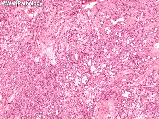
Differential Diagnosis of Sertoli cell tumor (SCT) of the Testis : SCT should be distinguished from: Seminoma: Rare seminomas may have tubular pattern. On the other hand, rare SCT may have diffuse sheet-like growth pattern, clear cytoplasm, and lymhocytic infiltrate mimicking seminoma. Features favoring SCT include: absence of germ cell neoplasia-in-situ; presence of areas with typical tubules; uniform bland nuclei; lack of prominent nucleoli; low mitotic activity; negativity for PLAP, OCT3/4 and SALL4. Androgen Insensitivity Syndrome: Patients with androgen insensitivity syndrome (also referred to as testicular feminization syndrome) may develop hamartomatous nodules composed of seminiferous tubules lined by Sertoli cells - referred to as Sertoli cell adenomas. They are usually multifocal and bilateral and closely resemble well-differentiated Sertoli cell tumors. In some cases, there is prominent basement membrane material deposited around individual tubules. Cryptorchid Testis: Aggregates of small seminiferous tubules lined by immature-appearing Sertoli cells are commonly found in cryptorchid testes. They are usually microscopic but sometimes may be large enough to be detected clinically or on testicular ultrasound. Leydig Cell Tumor (LCT): Features favoring LCT include: diffuse sheet-like growth pattern, bright eosinophilic cytoplasm, Reinke's crystalloids, consistent positivity for alpha-inhibin and CD99, and lack of nuclear beta-catenin positivity. Features favoring SCT include at least focal tubular differentiation, presence of multiple architectural patterns, pale cytoplasm, lack of Reinke's crystalloids and nuclear staining with beta-catenin. The image shows tubular, trabecular, and nested patterns in a Sertoli cell tumor.