
Rhabdoid Tumor

slide 32 of 84


Comments:
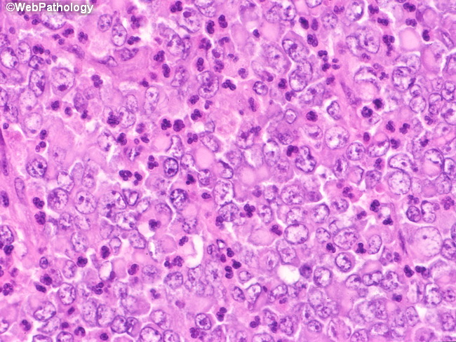
The characteristic intracytoplasmic eosinophilic globules seen in rhabdoid tumor are composed of intermediate filaments and are positive for vimentin and keratin. Muscle and neural markers are generally negative. Rhabdoid appearance is not specific for rhabdoid tumor of kidney and can be seen in other renal neoplasms, including mesoblastic nephroma, Wilms tumor, and renal cell carcinoma. In addition, "rhabdoid phenotype" may result from accumulation of intermediate filaments in a disparate group of neoplasms, including synovial sarcoma, desmoplastic small round cell tumor, rhabdomyosarcoma, malignant melanoma, and many other types of carcinomas.
slide 32 of 84