
Chondroblastic Osteosarcoma

slide 31 of 93


Comments:
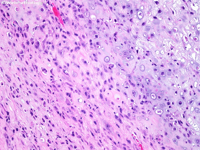
Cartilaginous differentiation is seen in the upper right corner. The tumor cells are present in lacunar spaces. The periphery of the cartilaginous area (lower left) is hypercellular with spindling of the tumor cells.
slide 31 of 93