
Borderline Brenner Tumor

slide 30 of 56


Comments:
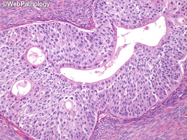
The image shows sharply-defined nests composed of cells resembling mature transitional epithelium (urothelium) within a dense fibromatous stroma. Some of the nests show central cystic spaces which are lined by cuboidal or columnar epithelium. The immunohistochemical profile of borderline Brenner tumors is similar to their benign counterparts. They are positive for CK7, p63, GATA3, uroplakin, thrombomodulin, EMA, CEA and S-100 protein. They are usually negative or only weakly positive for CK20 and p53.
slide 30 of 56