
Hairy Cell Leukemia : Bone Marrow


Comments:
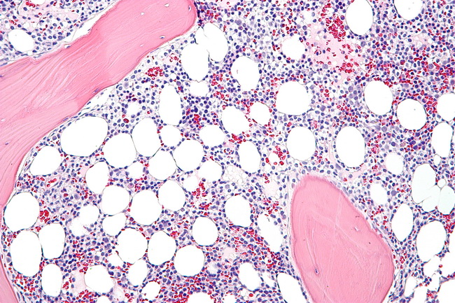
In HCL, the bone marrow biopsies are generally hypercellular and show diffuse sheets of hairy cells. Unlike other small B-cell lymphomas, well-defined cellular aggregates are not seen. In early stages, the bone marrow may even be hypocellular (and mimic aplastic anemia) and the leukemic cells may not be readily apparent on routine H&E stains. The hairy cells appear monotonous with oval nuclei which are surrounded by abundant clear cytoplasm creating a fried egg appearance. Hairy projections may become apparent with DBA.44 immunostain. The tumor cells may have spindled morphology in some cases.The normal hematopoietic elements (especially the myeloid lineage) are reduced and may show dysplastic changes mimicking myelodysplastic syndrome. Plasma cells and mast cells may be slightly increased. There is significant reticulin fibrosis in marrows affected by hairy cell leukemia.Examination of bone marrow is not essential for the diagnosis of HCL. The diagnosis can be rendered in most cases by evaluating peripheral blood smear alone on the basis of characteristic morphology and the immunophenotypic findings. However, it is useful to know the extent of bone marrow replacement by leukemic cells and it provides a baseline for assessing treatment response. A good cellular bone marrow aspirate is often difficult to obtain due to the presence of reticulin fibrosis, making it essential to get a proper bone marrow core biopsy. "Hairy cell leukemia - high mag" by Nephron - Own work. Licensed under CC BY-SA 3.0 via Wikimedia Commons.