
Aneurysmal Bone Cyst : Imaging - Case 1


Comments:
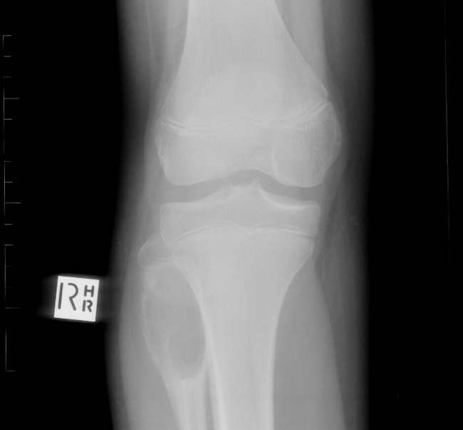
Aneurysmal Bone Cyst (ABC) - Imaging: ABC appears on radiographs as an expansile, multiloculated, lytic lesion with well-defined margins, located eccentrically in the metaphysis of long bones. There is marked thinning or even complete destruction of the cortex with tumor extension into soft tissues. Most lesions are surrounded by a thin shell of reactive bone. Less frequently, ABC is located on the bone surface and appears to arise from the cortex or the periosteum. This plain radiograph (frontal view) demonstrates a lytic expansile lesion involving the neck of right fibula that was diagnosed as ABC. Next six images (4-9) are from the same case and show the appearance of ABC on various imaging modalities. Case courtesy of Dr Alexandra Stanislavsky, Radiopaedia.org. From the case rID: 14333